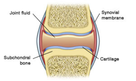
Normal Joint Structure
These are the Five (5) Main Components of a Joint
The Cartilage
Cartilage is a tissue that forms the lining of the bone ends. It has a pearlescent appearance and its thickness varies from one joint to another. Thus it is the kneecap that has the thickest cartilage: more than 5 millimeters. It is also worth knowing that men's cartilage is generally thicker than that of women.
The role of cartilage is to allow perfect sliding between the bones since the frictional forces are less than that of an ice skater sliding on ice. It is also essential for cushioning and distributing pressure on the bones thanks to its properties of elasticity and resistance.
Cartilage is a living tissue, in perpetual renewal, even among older people: it renews itself completely around every 3 months. Hence, microscopic cartilage fragments constantly break away from the cartilage and end up in the joint cavity (where they are then eliminated by the synovial membrane).
At microscopic level, it is composed of two components:
• On one hand, a matrix consisting of a gel rich in water and molecules called proteoglycans held tightly together in the meshes of the collagen fiber network. Proteoglycans provide the elasticity of the cartilage while the collagen fibers give it resistance to compressive forces.
• On the other hand, cells called Chondrocytes, whose role is fundamental because they have the dual ability to renew, but also to destroy the matrix.
• On the other hand, cells called Chondrocytes, whose role is fundamental because they have the dual ability to renew, but also to destroy the matrix.
The cartilage tissue has no blood vessel and it is not innervated.
The Joint (Synovial) Fluid
Joint fluid also called Synovial Fluid is secreted by the synovial membrane. It is present in all joints in small quantities (1 to 2 ml in the knee for example). Its role is to lubricate the joint. It ensures perfect sliding between the bone ends.
In its normal state it is a clear, transparent fluid with a particular viscosity. The latter is due to the amount of Hyaluronic Acid present in the joint.
In its normal state it is a clear, transparent fluid with a particular viscosity. The latter is due to the amount of Hyaluronic Acid present in the joint.
The Articular Synovial Capsule and Membrane
The articular synovial capsule and membrane are closely linked since the synovial membrane coats the inside of the capsule, a fibrous covering surrounding the joint.
The synovial membrane is a vascularized and innervated tissue. It is in the form of a smooth transparent membrane covered with small blood vessels. in its normal state it has folds called villi.
Its main role is to secrete the Hyaluronic Acid of the synovial fluid, a true lubricant of the joint. The synovial membrane also ensures "emptying" of cartilage debris found in the joint cavity. This is thanks to its blood vessels that bring oxygen and nutrients, including glucose, essential for the life of the cartilage.
At microscopic level, the synovial membrane is composed of cells called synoviocytes. It is these cells that perform the functions mentioned above. Lastly, these cells also have the ability to produce enzymes that can destroy the cartilage matrix.
Illustration and Information courtesy of Arthrolink.com / The Osteoarthritis Website
The Importance of the Articular (Joint) Cartilage’s Extracellular Matrix (ECM)
for the Entire Joint Structure
Articular (Joint) Cartilage is a hydrated, avascular tissue. Its principal function is to provide a smooth, lubricated surface for articulation and to facilitate the transmission of loads with a low frictional coefficient. The mechanical behavior of this tissue depends on the interaction of its fluid and solid components. The unique and complex structure of Articular Cartilage continues to make its treatment and repair a significant challenge.
It is primarily composed of Extracellular Matrix (ECM), which is a network of two major classes of biomolecules, namely : non-fiber Glycosaminoglycans (GAGs)(such as Hyaluronic Acid, Aggrecan/Chondroitin Sulfate Proteoglycan, etc.), interacting with Fibrous Proteins which include Collagen, Elastin, etc. All of these components are synthesized, maintained and secreted by the cells of Cartilage known as Chondrocytes.
The Chondrocyte is the resident cell type in Articular Cartilage. Chondrocytes are highly specialized, metabolically active cells that play a unique role in the development, maintenance, and repair of the ECM. Chondrocytes originate from mesenchymal stem cells and constitute only about 2% of the total volume of Articular Cartilage. Along with Collagen fiber ultrastructure and ECM, Chondrocytes contribute to the various zones of Articular Cartilage - the superficial zone, the middle zone, the deep zone, and the calcified zone
Water is the most abundant component of Articular Cartilage, contributing up to 80% of its wet weight. Unlike most tissues, Articular Cartilage does not have blood vessels, nerves, or lymphatics. Together, the above described components help to retain water within the ECM, which is critical to maintain its unique mechanical properties. The flow of water through the Cartilage and across the articular surface helps to transport and distribute nutrients to the Chondrocytes, in addition to providing lubrication.
Collagen is the most abundant structural macromolecule in ECM, and it makes up about 60% of the dry weight of cartilage. Type II collagen represents 90% to 95% of the Collagen in ECM and forms fibrils and fibers intertwined with proteoglycan aggregates.
Proteoglycans are heavily glycosolated protein monomers. In articular cartilage, they represent the second-largest group of macromolecules in the ECM and account for 10% to 15% of the wet weight. The largest in size and the most abundant by weight is aggrecan, a proteoglycan that possesses more than 100 chondroitin sulfate and keratin sulfate chains. Aggrecan/Chondroitin Sulfate is characterized by its ability to interact with Hyaluronan (Hyaluronic Acid) to form large proteoglycan aggregates via link proteins. Aggrecan occupies the interfibrillar space of the cartilage ECM and provides cartilage with its osmotic properties, which are critical to its ability to resist compressive loads.
The degradation (loss or substantial deficiency) of any of these major macromolecular components of the Extracellular Matrix (ECM) from the Cartilage results in serious impairment of joint function and in a major cause of Osteoarthritis (OA).
During normal cartilage turnover (metabolism) in healthy articular joints, ECM production balances ECM breakdown, thereby ensuring the continuous renewal of this critical joint-cushioning tissue. However under pathological (disease) conditions, ECM synthesis cannot keep pace with degradation and a loss of the structural integrity of the Articular Cartilage results.
Joint motion and load are important to maintain normal articular cartilage structure and function. Inactivity of the joint has also been shown to lead to the degradation of cartilage. Regular joint movement and dynamic load is important for the maintenance of healthy articular cartilage metabolism. The development of disease such as Osteoarthritis(OA) is associated with dramatic changes in cartilage metabolism. This occurs when there is a physiological imbalance of degradation and synthesis deficiency by the Chondrocytes.
Due to its foremost important position in the Articular (Joint) Cartilage, we must seek to sustain the vital function of the Chondrocytes by meeting their nutritional needs with the recognized best Chondroprotective natural substances available in the market.
The Importance of Synovial Fluid to Joint Health
The synovium is a membrane surrounding each joint and is responsible for the production of a thick, slippery fluid. Moving into the cartilage when a joint is at rest and out to the joint capsule during activity, synovial fluid helps ensure smooth and easy body movement. It lubricates and nourishes joints, making it a necessary component for optimal joint health.
Hyaluronic Acid is a vital component of synovial fluid. Found in the highest concentrations in fluids lining the joints and eyes, hyaluronic acid (HA) is a vital component of synovial fluid. The presence of this important acid creates a cushion for joint cartilage, reducing the friction of bone-on-bone contact.
People with diseases like arthritis experience inflammation within the synovium, which causes pain and stiffness in the joint and limited mobility. Some of the best supplements for joints include Hyaluronic Acid in the ingredients.
In healthy joints, hyaluronic acid in synovial fluid is continuously broken down and replaced. In osteoarthritic joints, breakdown happens faster than production, which results in watery synovial fluid that doesn't function properly. Cartilage in the joint eventually erodes, causing pain, stiffness, and swelling.
‘CHONDROPROTECTION’
via the utilization of a ‘Chondroprotective’ compound such as the synergistic combination of select functional ingredients that you find exclusively in our proprietary Mobile and Flexible All-In-One Joint Health Formula TM - Is Arguably, The Key to Enhance The Health of The Entire Joint Structure (Chondrocytes, Cartilage, Subchondral Bone,Synovial Fluid and Membrane & Articular Cartilage), thus preserving normal joint function by effectively interfering with the anatomical progression of Osteoarthritis (OA).
